Periodontology
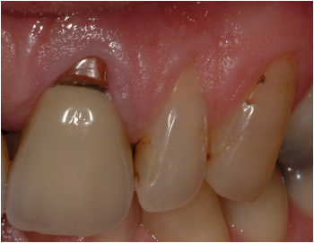
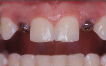
Before

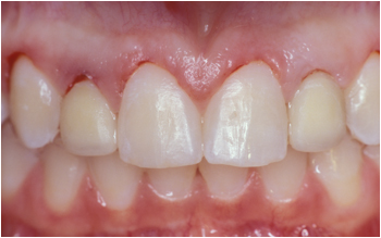
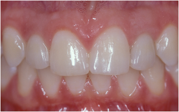
After
Crown Lengthening Procedure
The benefits of crown lengthening procedures are both functional and esthetic. As one of the most commonly used procedures in periodontics, it is indicated when insufficient crown length prevents adequate retention to the future restoration. This procedure will adjust the bone and gingival levels to expose more tooth structure. It will improve the health and appearance of the tissue around the future prosthesis.
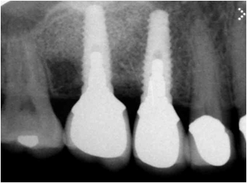
Dental Implants
Dental implants are the best available solution for the problem of missing teeth. Made out of titanium and shaped to replace the root of the missing tooth, they offer several advantages over conventional crown and bridgework. They conserve the integrity of the adjacent teeth, offer a longer lifespan, prevent further bone loss and provide excellent aesthetics.
Replacing a missing tooth with a dental implant require several steps involving your periodontist as well as your general dentist. The process begins with a thorough comprehensive examination of the condition and quantity of available gum and bone, along with special radiographs to locate the position of vital structures (nerves, sinus cavity etc). Treatment with dental implants usually starts with the surgical placement. Access to the bone is necessary to insert the implant before the gums are replaced and secured in their original position. A healing period of 3 to 5 months will follow depending on where the implant is placed, after which the restoring dentist will complete the treatment by connecting the final crown to the implant.
Sinus Augmentation
The upper back teeth are usually situated immediately beneath the sinus. The bone height between the floor of the sinus and the root of the molar is often thin or absent.
If bone gets lost in that area due to tooth loss or periodontal disease, sinus augmentation will be indicated to regenerate enough bone in height and width for the placement of dental implants.
Surgical exposure of the deficient area is necessary to raise the sinus floor. The gums are lifted away from the ridge and the sinus cavity is accessed from the side through a trapdoor-like preparation. The space underneath will then be filled with bone graft material and the gums are repositioned and secured with sutures.
The surgical area is allowed to heal for 6 to 12 months after which the implant is placed. In some cases, implants can be placed simultaneously with the sinus augmentation procedure.
Site Development/Ridge Augmentation
Height and width of the available bone are key to the success of dental implants. Tooth loss can often leave deformities and/or concavities in the upper or lower jawbone that complicate placement of implants.
In such cases bone augmentation is indicated to develop an adequate implant site. The volume needed will be provided by grafting bone or bone substitute in the deficient area. Surgical exposure of the area is necessary, the gum is lifted away from the ridge, the defect is then filled with bone after which the gum is repositioned and secured with sutures.
Healing will take from 4 to 12 month and the area will be ready for implant placement. In some cases, implants can be placed simultaneously with the augmentation procedure.
Aesthetic Periodontal Procedures
Connective Tissue Graft
Gingival recession can lead to increased sensitivity of the tooth, increased incidences of root surface caries, impairment in oral hygiene measure and an unappealing look. Individuals that get recession tend to have thinner tissues and consequently are more vulnerable to developing these recessions. In addition, periodontal disease and/or excessive trauma could contribute to the development of recession.
A connective tissue graft is a gingival augmentation procedure that uses the patient’s own tissue (usually taken from the palate) to graft directly onto the recessed area. As a result, the graft will cover the denuded area, reduce further recession, and protect the tooth against decay.
Gingival Graft
The lack of adequate width of gingiva in conjunction with restorative procedures, orthodontic therapy, or poor oral hygiene could lead to the resorption of the gingival margin. The free gingival graft will provide an adequate zone of attached gingiva that will allow proper long-term maintenance. The tissue is usually harvested from the palate and placed onto the affected area.
Frenectomy
Frenectomy is the removal of the frenum, which is a fold in the gingiva that passes from the lip to the gingival margin. A high frenum insertion sometimes retracts the gingiva when the lip is stretched. This stretching and distention can be associated with spacing between teeth, mal-alignment, recession and impairment of proper hygiene.
The removal of this frenum is done under local anesthesia, the frenum base is separated from the gingiva and sutures are placed to close the wound.
Gingivectomy/Gingivoplasty
Gingivectomy or Gingivoplasty is the surgical removal and re-contouring of the gingiva in cases of excessive gingival display (gummy smile) or drug induced gingival overgrowth. This procedure will provide a cosmetic enhancement of the gingiva as well as improved access for proper hygiene and maintenance.
Treatment of Periodontal Disease
Treatment options for periodontal disease may vary depending on the extent and severity of tissue destruction. Usually it is divided into surgical and non-surgical treatment. The main objective of periodontal therapy is to eliminate all the disease causing bacteria and regenerate the supporting structures of the affected tooth.
Non-surgical pocket therapy, also called scaling and root planing, is the initial procedure that reduces the signs of infection. The result of this initial phase is a noticeable improvement in the clinical condition. This includes a significant reduction in swelling and bleeding as well as reduction in pocket depth.
Surgical pocket therapy is indicated in more advanced cases where scaling and root planing alone are not sufficient. If it is not possible to eradicate all the bacteria colonizing the deep pockets, visual surgical access is then necessary.
Types of surgical treatment commonly prescribed include:
Resective Pocket Reduction Procedures
Periodontal pockets are the result of the separation of the gum from the affected tooth. A space will develop where bacteria will colonize and infect the underlying supporting bone. Over time these pockets become deeper allowing more bacteria and more destruction. If too much bone is lost, the teeth will need to be extracted. The Periodontist measures the depth of the pocket and determines that a flap procedure is necessary. This includes the separation of the gum from the tooth under local anesthetic and the removal of the disease causing bacteria. The infected tissues are then removed before securing back the gums, usually with sutures.
Regenerative Procedures
These procedures could reverse the destructive process by regenerating lost bone and tissue. This includes the separation of the gum from the tooth under local anesthetic and the removal of the disease causing bacteria along with the infected tissues. Bone graft or growth stimulating proteins could be applied to help regenerate the bone. The gums are then repositioned and secured back in place with sutures.
Connective Tissue Graft
Gingival recession can lead to increased sensitivity of the tooth, increased incidences of root surface caries, impairment in oral hygiene measure and an unappealing look. Individuals that get recession tend to have thinner tissues and consequently are more vulnerable to developing these recessions. In addition, periodontal disease and/or excessive trauma could contribute to the development of recession.
A connective tissue graft is a gingival augmentation procedure that uses the patient’s own tissue (usually taken from the palate) to graft directly onto the recessed area. As a result, the graft will cover the denuded area, reduce further recession, and protect the tooth against decay.
Before
Before
After
Sinus Augmentation
The upper back teeth are usually situated immediately beneath the sinus. The bone height between the floor of the sinus and the root of the molar is often thin or absent.
If bone gets lost in that area due to tooth loss or periodontal disease, sinus augmentation will be indicated to regenerate enough bone in height and width for the placement of dental implants.
Surgical exposure of the deficient area is necessary to raise the sinus floor. The gums are lifted away from the ridge and the sinus cavity is accessed from the side through a trapdoor-like preparation. The space underneath will then be filled with bone graft material and the gums are repositioned and secured with sutures.
The surgical area is allowed to heal for 6 to 12 months after which the implant is placed. In some cases, implants can be placed simultaneously with the sinus augmentation procedure.
GET IN TOUCH
The Atrium Clinic, The Atrium, Blackpool Business Park, Blackpool, Cork.
T: 021 439 9056 E: [email protected]
request an appoinTment
Error: Contact form not found.